By Richard Dargan

Cardiac MRI is an effective means to study the structure and function of the heart without exposing the patient to ionizing radiation. However, standard steady state free procession (SSFP) cine acquisitions, the mainstay of the cardiac MR examination, are limited by long acquisition times, data segmentation over multiple heart beats and breath-hold requirements. Many patients who undergo the examination struggle to perform required breath holds in the claustrophobic environment of the scanner.
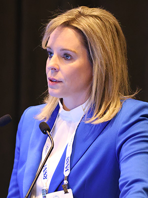
“These are patients who have cardiovascular diseases and cardiomyopathies,” said study lead author Melany Beth Atkins, MD, from Fairfax Radiological Consultants in Virginia. “So, we really need a way to acquire these SSFP images faster.”
Dr. Atkins and colleagues studied highly accelerated cine with deep learning image reconstruction to speed examination times without a loss of image quality. The 70 patients in the study underwent cardiac MR with SSFP and two deep learning-assisted techniques: one with three heartbeats per slice and one with a single heartbeat per slice.
Average total scan time for standard SSFP was 7.9 minutes, compared with only 2.7 minutes for the three heartbeat technique and 1.6 minutes for the single heartbeat. Scan time savings were 66% and 80%, respectively, with comparable image quality to the conventional SSFP cine images. The volumetric and function measurements demonstrated negligible differences between the three acquisition types.
This has been a huge breakthrough,” Dr. Atkins said. “The image quality is at least the same in most patients and even better in certain patients like those with an arrhythmia who can’t hold their breath.”
Dr. Atkins described several cases that illustrate the technology’s potential. In one, a 76-year-old man with claustrophobia had difficulty holding his breath during the examination, resulting in suboptimal SSFP images. The three heartbeat and single heartbeat images, in contrast, demonstrated left ventricular hypertrophy and associated findings excellently.
Between the two types of highly accelerated cine, the three-heartbeat model offers high spatial resolution but is not as fast as the single heartbeat model and still requires a breath-hold sequence. Respiratory triggering with bellows, a technique that uses biofeedback to collect data when diaphragm motion is minimal, can enable the procedure to be done without a breath hold. A future goal will be expanding the respiratory triggering capabilities in the three heartbeat model while maintaining its resolution.
According to Dr. Atkins, along with improved patient comfort and image quality, the shorter scan time reduction will also allow for additional patient throughput, successful exam completion and access to cardiac MR in locations with long patient backlogs. These benefits are among the reasons why she hopes to deploy the deep learning technology to other types of cardiac imaging at her facility.
“The goal is to expand this to the whole gamut of cardiac imaging and be able to image the majority of our patients in under 20 minutes without them having to hold their breath,” she said.
Access the presentation, “Evaluation of Highly Accelerated CINE with Deep Learning Image Reconstruction to Decrease Scan Time While Maintaining Image Quality and Accurate Volumetric Analysis,” (M1-SSCA03-1) on demand at RSNA.org/MeetingCentral.
© 2024 RSNA.
The RSNA 2024 Daily Bulletin is the official publication of the 110th Scientific Assembly and Annual Meeting of the Radiological Society of North America. Published online Sunday, December 1 — Friday, December 6.
The RSNA 2024 Daily Bulletin is owned and published by the Radiological Society of North America, Inc., 820 Jorie Blvd., Suite 200, Oak Brook, IL 60523.