By Jennie McKee
Chronological age is an imperfect measure of health and longevity, noted Matthew Lee, MD, an assistant professor in the Department of Radiology at the University of Wisconsin School of Medicine and Public Health in Madison.
“Biological age (BA), on the other hand, is a potentially useful concept that encapsulates not only the passage of time, but also the collective impact of genetic predisposition, lifestyle habits and disease processes on an individual’s health,” he said.
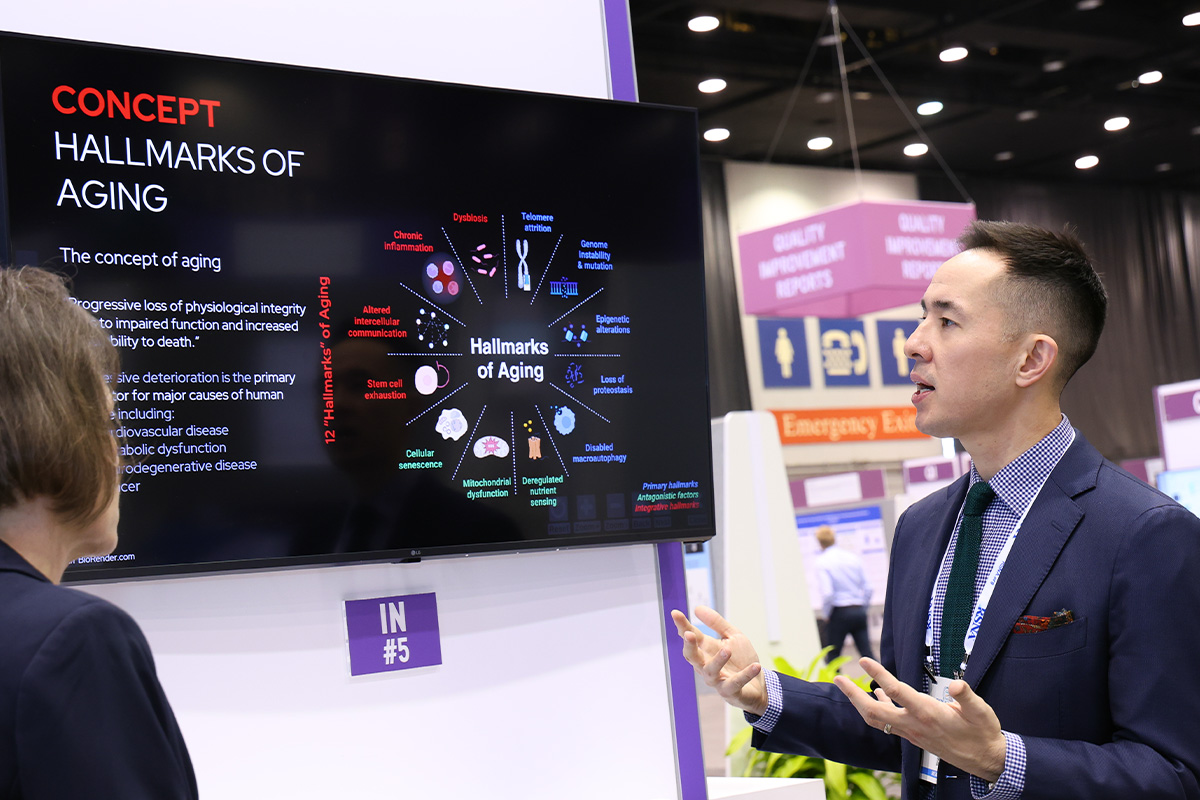
In his education exhibit presented Tuesday, Dr. Lee reviewed basic concepts of aging and the existing BA landscape. He also discussed tools for BA estimation as well as the role and impact of radiology and image-based BA estimation for aging research and clinical application.

Existing BA models have focused on clinical, molecular and cellular ‘omics’ biomarkers, Dr. Lee asserted.
“Imaging biomarkers have received less attention but may offer valuable insights into the ‘phenotypic’ cumulative macroscopic effects of aging that currently go underutilized,” he said.
Unlike the molecular and cellular-level "frailomics," as well as clinical measures used in existing BA models, imaging biomarkers are accessible, reproducible and may also better capture the big picture phenotypic tissue-level effects of aging, Dr. Lee noted.
With technological advances in computing power and the advent of fully automated tools, imaging biomarkers can be used as predictive, prognostic or treatment biomarkers.
“Machine learning (ML) techniques have been essential in distilling large volume omics data,” Dr. Lee said. “ML is ideal for big data and estimating BA from ‘non-conventional’ parameters, such as imaging, although there is no specific ML technique dedicated to BA estimation.”
ML methods have a black box effect. The lack of model interpretability remains a major challenge for deep learning-based aging clocks, Dr. Lee noted.
According to Dr. Lee, a benefit of explainable AI tools is that they enhance confidence in model prediction by being inherently more transparent and understandable.
“At our institution,” Dr. Lee said, “work in opportunistic imaging by Perry Pickhardt, MD, and colleagues has shown that CT can provide objective, reproducible measures of abdominal muscle, fat, aortic calcification, organs and bone associated with age-related disease.”
Dr. Lee added that these predictive CT biomarkers of survival and adverse clinical events such as cardiovascular events and fragility fracture show promise as quantitative biomarkers of aging that can be used to estimate BA.
The explainable AI-driven CT biomarkers can be used in a combined “phenotypic” model that can be retrospectively applied to existing scans without the need for additional testing or patient dose.
Using imaging biomarkers for BA estimation could help identify patients at risk for developing age-related diseases or patients who already have them and improve existing clinical tools for risk prediction.
“Continued work to improve image-based methods for BA estimation could meaningfully impact the lives of our patients,” Dr. Lee said, noting that this work has garnered interest from colleagues outside of radiology at his institution.
According to Dr. Lee, future research efforts may include large-scale validation of image-based BA age models using AI tools in larger, more diverse patient populations, using clinical scan data, comparing image-based models to other ‘omics’ BA models, and the creation of models based on multisystem tissue assessment.
Access the education exhibit, “CT Imaging Biomarkers for Phenotypic Biological Aging: Overview and Comparison with Other Radiological and Non-Radiological Approaches,” (INEE-5) on demand at RSNA.org/MeetingCentral.
© 2024 RSNA.
The RSNA 2024 Daily Bulletin is the official publication of the 110th Scientific Assembly and Annual Meeting of the Radiological Society of North America. Published online Sunday, December 1 — Friday, December 6.
The RSNA 2024 Daily Bulletin is owned and published by the Radiological Society of North America, Inc., 820 Jorie Blvd., Suite 200, Oak Brook, IL 60523.